 Scleral Buckling - procedure, recovery, removal, pain, complications, time, infection, medication
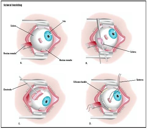
Scleral pande Definition Scleral pandeo is a surgical procedure in which a piece of silicone plastic or sponge is sewn on the sclera on the site of a retina tear push the sclera into the retina tear. The buckle holds the retina against the sclera until the healing seals the tear. Also prevents the liquid filtration that could cause more retinal detachment. Purpose The scleral pande is used to re-assembly the retina if the rest is very large Or if the tear is in a place. It is also used to seal breaks in the Retina. Demographics Retinian detachment occurs in 25,000 Americans each year. Patients suffering of retinal detachments are commonly near the sight, have had eye surgery, experienced eye trauma, or having a family history of retina Highlights. Retinian detachments are also common after the elimination of cataracts. White men have a higher risk, such as people who are middle-aged or Major. Patients who have already had a retinal detachment also have a greater opportunity for another detachment. Some conditions, such as diabetes or Coats disease in children, make people more susceptible to retinal detachments. Description The scleral pandeo is made in a under general or local anesthesia. Immediately before the procedure, patients are given eye drops to dilate the student to allow better access the eye. He's given a local anesthesia. After the eye is numb, the surgeon cuts the eye membrane, exposing the sclera. If bleeding or swelling blocks the surgeon's view of retinal detachment or hole, he or she can perform a vitectomy before the scleral pande. Vitretomy is necessary only in cases where the surgeon's opinion the damage is hindered. The surgeon makes two incisions in the sclera, one for a light probe and the other for cutting instruments vacuum. The surgeon uses a small device, similar to the guillotine to remove the vitreus, which then replaces with saline. After the removal, The surgeon can inject air or gas to keep the retina in place. Then the surgeon is able to see the retina, he or she will perform one of two peer procedures. In a scleral pandemic procedure, one of the rectum of the eye muscles are cut to get access to sclera (A). The sclera is cut open (B), and an electrode is applied to the area retinal detachment (C). A silicone buckle gets in place under the muscles of the rectum (D), and the severe muscle is repaired. After the surgeon has performed laser or cryopexia photocoagulation, he or indenta the affected area of sclera with silicone. The silicone, either in the form of sponge or buckle, close the tear and reduce the The circumference of the eyes. This reduction prevents further pulling and separation from the vitreous. Depending on the gravity of the detachment or hole, a buckle can be placed around the entire eyeball. When the buckle is in place, the surgeon can drain subretinal fluid that I could interfere with the retina readjustment. After the liquid is the surgeon will stitch the buckle in place and then cover it. with the conjunctive. The surgeon then inserts an antibiotic (drops or ointment) in the affected eye and patch it. For less severe detachments, the surgeon may choose a temporary buckle that will be eliminated later. Usually, however, the buckle remains in place for the patient's life. It It doesn't interfere with vision. Scleral buckles in babies, however, needs to be removed while the eyeball grows. Diagnosis/Preparation Retinian detachment is considered an emergency situation. In the case of a Acute start-up detachment, the time it takes to repair detachment, less chances of successful reassignment. Normally the patient sees floating stains and experiences peripheral loss of visual field. Patients commonly describe the loss of vision as having someone pulling the shadow over their eyes. In extreme cases, patients may lose sight completely. An ophthalmologist or optometrist will take a complete medical history, including the family history of retinal detachment and any recent eyepiece trauma. In addition to a general eye exam, including a examination of the cut lamp, macula examination and lens assessment, doctors can perform the following tests to determine the degree of retina detachment: Small breakages in the retina do not require surgery, but patients with Acute start-up detachment requires reconnection within 24 to 48 hours. Chronicle Retinal detachments must be repaired within a week. Because scleral pandeo is usually an emergency procedure, there is no Long-term preparation. Patients should fast at least six hours before surgery. Aftercare Immediately after surgery, patients will need food and food help walking. Some patients should remain in hospital for several days. Many However, scleral pandemic procedures are performed on an outpatient basis. After hospital release, patients should avoid heavy lifting or arduous that could increase intraocular pressure. Quick eye movements must also be avoided; reading may be prohibited until the surgeon gives permission. Sunglasses should be used during the day and a patch of eyes at night. Pain and a feeling of scratch, as well as redness in the eye can also occur after surgery. Ice packets can be applied if the conjunctiva is swollen. Patients may take pain medications, but should consult with your doctor before taking any free-sale medications. Excessive pain, swelling, bleeding, eye discharge or decrease vision is not normal, and should be reported immediately to the doctor. If a vitretomy is performed in conjunction with the scleral pande, Patients should sleep with their head elevated. They should also avoid the air travel until the air bubble is absorbed. After scleral greed, patients will use dilation, antibiotic or eye drops corticosteroids for up to six weeks to decrease inflammation and the possibility of infection. The best visual acuity cannot be determined to at least six to eight weeks after surgery. Driving can be prohibited or restricted while the vision stabilizes. In the post-operative period of six to eight weeks visit, doctors determine whether the patient needs remedial lenses or stronger prescription lenses. The restoration of the complete vision depends on the location and gravity of the detachment. Risks Complications are rare but can be severe. In some cases, patients lose seen in the affected eye or lose the entire eye. The scar tissue, even the pre-existing scar tissue, can interfere with the scar tissue retina readjustment and scleral pande procedure may have to be repeated. Fear, along with infection, is the most common complication. Other possible but rare complications include: Patients can also be more watched after the procedure. In some instances, although the retina rettaches, the vision is not restored. Normal results National Institutes of Health report that scleral turmoil has a 85-90% success rate. The restored vision depends to a large extent on the location and extension of the detachment, and duration of time before The detachment was repaired. Patients with peripheral detachment have a recovery faster then those patients whose detachment was located in the Macula. The longer the patient waits to repair the detachment, the worse the prognosis. Morbidity and mortality rates The risk of mortality and loss of vision depends on the cause of retina detachment. Patients with Marfan syndrome, preeclampsia and diabetes, for example, is more at risk during scleral slimming procedure that a patient in relatively good health. The risk of surgery also increases with the use of general anesthesia. However, scleral, is considered a safe and successful procedure. Severe infections that remain untreated can cause vision loss, but following the prescribed regimen of eye drops and follow-up treatment by the doctor greatly minimizes this risk. Alternatives Vitretomy is sometimes done only to treat retinal detachments. Laser photocoagulation and cryopexia can also be used to treat less severe tears. The most common alternative, however, is tyre retinopexy, which is used when the tear is at the top of the eye. The surgeon uses chryopexia to freeze the area around the tear, then it is removed a small amount of liquid. When the liquid is drained and the eye softens, the surgeon injects a gas bubble into the vitreous cavity. Like gas bubble expands, seals the retina tear by pushing the retina against the choroid. Eventually, the bubble will be absorbed. The patient must remain in a given position at least a few days after surgery while the bubble helps to seal the hole. Pneumatic retinopexy is also not as successful as scleral pandeo. Complications include recurrent retinal detachments and the possibility of gas being put down the retina. Resources books Buettner, Helmut, M.D., editor. Mayo Clinic in Vision and Ojo Cheers. Rochester, MN: Mayo Clinic Health Information, 2002. Cassel, Gary H., M.D., Michael D., Billig, O.D. and Harry G. Randall, M.D. The Book Eye: A Complete Guide to Eye Disorders and Health. Baltimore, MD: Johns Hopkins University Press, 1998. All you need to know about medical treatments , edited by Stephen Daly. Springhouse, PA: Springhouse Corp., 1996. Sardgena, Jill, et al. The Encyclopaedia of Ceguera and Vision, 2nd edition. New York, NY: File data, Inc. 2002. organizations American Academy of Ophthalmology. PO Box 7424, San Francisco, CA 94120-7424. (415) 561-8500. . American Board of Ophthalmology. 111 Presidential Boulevard, Suite 241, Bala Cynwyd, PA 19004-1075. (610) 664-1175. info@abop.org. . National Eye Institute. 2020 Vision Place, Bethesda, MD 20892-3655. (301) 496-5248. . others Handbook of Ocular Disease Management: Retinal Detachment Review of Ophthalmology [quoted on 21 April 2003]. . "Retinian detachment." [quoted on 12 April 2003]. . "Retinal detachment repair." [cited May 1, 2003]. . Wu, Lihteh, M.D. "Retina detachment, exudative." . 28 June 2001 [quoted 1 May 2003]. . Mary Bekker Who does the contract and where is it reported? The scleral pande can be made by a general ophthalmologist, an M.D. that specializes in the treatment of the eye. Even more specialized ophthalmologists, vitreo-retina surgeons who specialize in diseases the retina, can be called for serious cases. Surgery is usually performed in hospital settings. Because of the delicacy of the procedure, sometimes a night hospital stay is required. Less severe retinal detachments may be treated in an outpatient base in surgery centers. QUESTIONS RELATING TO THE DOCUTOR User Contributions:Comment on this article, ask questions or add new information on this topic:
COVID-19 Update We are experiencing a volume of extremely high calls related to the interest of the COVID-19 vaccine. Please understand that our phone lines should be clear for urgent medical care needs. We cannot accept phone calls to schedule COVID-19 vaccines at this time. When this changes, we will update this website. Please know that our supply of vaccines is extremely small. Read all . Silence Silence Popular Silence SearchesScleral Buckling What is the scleral pande? Scleral pandeo is a type of eye surgery to correct a careless retina and restore vision. The retina is a layer of cells in the back of the eye. These cells use light to send visual information to your brain. Retina detachment occurs when part of your retina is separated from the rest of your retina and eye. When that happens, your retina doesn't work normally, and the vision is lost in all or part of your retina. If not treated promptly, this can cause permanent vision loss. Your surgeon may perform a scleral balance under local or general anesthesia. During the scleral balance, your eye surgeon will expose your eyeball. He or she can use a freezing instrument to help seal their retina together. After that, your surgeon can use a small device, a scleral buckle, to keep your retina in place. Why would I need scleral pande? Certain factors make it more likely that you will have a retina detachment. These include:Most of the time, retinal detachment occurs spontaneously, but sometimes eye trauma can also cause it. If you have retinal detachment, you will probably need some kind of procedure or surgery. You could have an increase in floats in your eye. These look like small spots or cobwebs floating in your field of vision. These floaters can be so dense that they hurt their vision. You can also experience light flashes in your eye or curtain over your field of vision. If you have these symptoms, you may need emergency surgery or a procedure to reconnect the retina. This can restore vision. Eye care providers sometimes treat retinal detachment with a less invasive procedure called pneumatic retinopexy. This procedure cannot treat all kinds of tears to your retina. However, if you have a complicated retina tear, you may also need another surgery called vitretomy. All of these techniques can successfully repair a retina that is uncovered. Ask your eye care professional about the benefits and risks of all your treatment options. What are the risks of scleral? Most people do well with scleral pande surgery, but complications sometimes happen. Your risks may depend on your age, medical conditions, and specific characteristics of your retinal detachment. The risks of the procedure are: There is also a risk that a retinal detachment will return and that you will need another surgery. How do I prepare for the scleral pandeo? Ask your eye care provider what you need to do to prepare for scleral pande surgery. Ask if you need to stop taking any medication before the procedure. You'll have to avoid eating something after midnight the day before surgery. Your healthcare provider may want to use special tools to shine a light in your eye and examine your retina. You may need your eyes to be dilated for your eye exam. You can also have an ultrasound of your eye, which helps your eye care provider to see the retina detachment. What happens during the scleral pande? Talk to your eye surgeon about what to expect during your surgery. Details can vary a bit. You probably made scleral pandemics in a hospital O.R. In general, during the procedure: What happens after the scleral? Ask your eye care provider what to expect after your surgery. In most cases, you will be able to go home the same day. He's planning on taking you home from the procedure. Be sure to follow your healthcare provider's instructions on eye care. You may need to take eye drops with antibiotics to help prevent infection. Your eye may be a little pained after the procedure, but you should be able to take medications for the over-the-counter pain. You may need to use an eye patch for a day or so. You will need close follow-up care with your surgeon to see if the procedure was effective. You may have a scheduled appointment the day after the procedure. Be sure to tell your surgeon right away if you have a decreasing vision, or increase pain, or swelling around your eye. If the procedure does not work, you may need additional surgery. Next Steps Before accepting the test or procedure, be sure to know: Specializing in:In another Johns Hopkins Hospital:Find additional treatment centers in:RelatedRelatedVitrectomyPneumatic RetinopexyThe best way to get a vision of age Related IssuesHealth

Series of photographs of a patient after scleral buckling using tissue... | Download Scientific Diagram
Scleral Buckle Surgery: Costs & Preparation Guide | NVISION Eye Centers
Removal of Hydrogel Scleral Buckles - Retina Today
Retinal detachment and Scleral Buckle eye surgery - Album on Imgur
Haunted By History
Surgery for Retinal Detachment: What to Expect at Home
Recovery photos | Eye surgery, Surgery, Recovery
Scleral Buckle Surgery for Retinal Detachment – Retina Specialist | Fairfax, Virginia | Retinal Diseases
Detached retina surgery recovery
Infectious conjunctivitis caused by Pseudomonasaeruginosa in infected and extrused scleral buckles | BMJ Case Reports
scleral buckle | joyceholmes
Storm Anesthesia - Eye treatment modalities
My Eye Injury: A physical and emotional battle - Why Things Hurt
Retinal detachment and Scleral Buckle eye surgery - Album on Imgur
One week after combine procedure of vitrectomy and scleral buckle;... | Download Scientific Diagram
What Is the Recovery Time After Detached Retina Surgery? | NVISION Eye Centers
Retinal Detachment Repair
Scleral Buckling | Johns Hopkins Medicine
Scleral Buckling: Surgery, Procedure Details, and Recovery Time
dilated pupil after eye surgery | joyceholmes
Scleral Buckle vs. Vitrectomy – Retina Specialist | Fairfax, Virginia | Retinal Diseases
Dispensing case study: Balancing vision requirements with additional concerns - Spectrum ANZ
Scleral Buckle – deerutty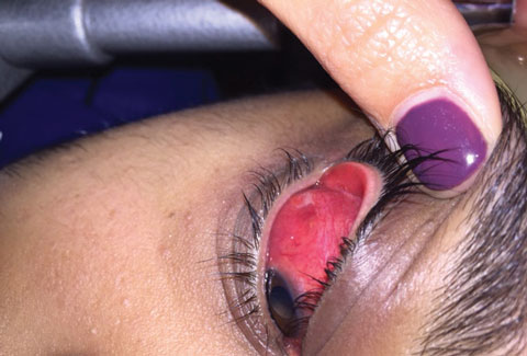
Haunted By History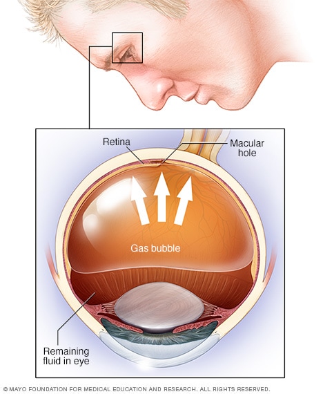
Retinal detachment - Diagnosis and treatment - Mayo Clinic
How Long Does a Scleral Buckle Last?
My Eye Injury: A physical and emotional battle - Why Things Hurt
Retinopathy of Prematurity (for Parents) - Nemours KidsHealth
PDF) A rare case of scleral buckle infection with Curvularia species
Scleral Buckle: How It Can Help | Dean McGee Eye Institute
Retinal detachment and Scleral Buckle eye surgery - Album on Imgur![Full text] Scleral buckling in the management of rhegmatogenous retinal detachmen | OPTH Full text] Scleral buckling in the management of rhegmatogenous retinal detachmen | OPTH](https://www.dovepress.com/cr_data/article_submission_image/s153000/153717/153717-figure-790.jpg)
Full text] Scleral buckling in the management of rhegmatogenous retinal detachmen | OPTH
LOOKING THROUGH THE BUBBLE — Spectacle Optometry
Words of Wisdom From The Elder » Blog Archive » The Road To Recovery
Understanding Scleral Buckling Surgery
Scleral Buckling: Surgery, Procedure Details, and Recovery Time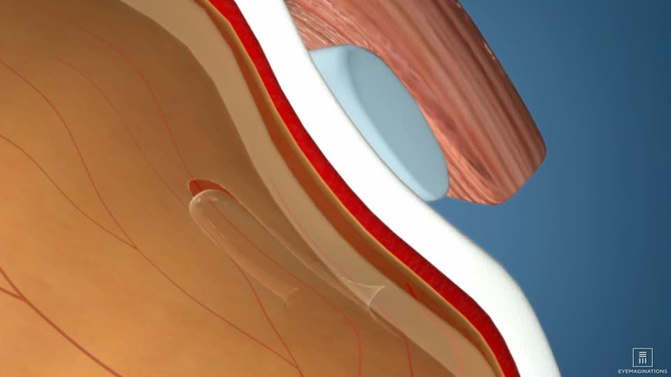
Cryo-Buckle Surgery for Retinal Detachment | Retina Doctor Melbourne
Eye Surgery and Female Bonding – deerutty
Retinal Detachment: Diagnosis and Treatment - American Academy of Ophthalmology
Retinal Surgery Asheville | Retinal Detachment Hendersonville
 Scleral Buckling - procedure, recovery, removal, pain, complications, time, infection, medication


![Full text] Scleral buckling in the management of rhegmatogenous retinal detachmen | OPTH Full text] Scleral buckling in the management of rhegmatogenous retinal detachmen | OPTH](https://www.dovepress.com/cr_data/article_submission_image/s153000/153717/153717-figure-790.jpg)


Posting Komentar untuk "scleral buckle recovery time"